ASSIST
Automated Structure-based Segmentation method for the Identification and Staging of Tumor aggressiveness
Today, histopathological diagnosis increasingly requires accurate quantification, classification, and structured documentation of findings. ASSIST‘s artificial intelligence supports pathologists in the analysis of digitized slides, providing advanced segmentation, classification, and quantification tools.
Histopathological diagnosis is becoming increasingly challenging and complex
Histopathological diagnosis is not limited to the observation of samples, but increasingly requires accurate quantification, classification, and structured documentation of findings. The growing complexity of oncology cases and the increase in the number of biomarkers to be evaluated place a considerable workload on pathologists, who must ensure accuracy, traceability, and speed in reporting. In this context, traditional manual methods are often slow, subjective, and difficult to scale, negatively impacting the overall efficiency of the department.
ASSIST alongside pathologists
ASSIST* is an artificial intelligence-based product designed to support pathologists in the analysis of digitized slides, providing advanced segmentation, classification, and quantification tools.
The device is part of a complex diagnostic process, in which the identification and characterization of histological structures is fundamental to the diagnostic process. ASSIST is designed to increase diagnostic accuracy, reduce variability, and speed up reporting, offering a complete set of automated tools that remain under the control of the professional.
Key features include:
- Segmentation, classification and characterization of histological structures
- Objective quantification
- Reporting and clinical traceability
*Device undergoing certification according to Regulation (EU) 2017/746 (IVDR).
ASSIST Prostate
ASSIST* Prostate uses AI models to locate and characterize adenocarcinoma in digital images of prostatic tissue with hematoxylin and eosin staining.
- It automatically segments tumoral areas, classifying them into Gleason pattern 3, 4 and 5
- It provides segmentations, excluding non-representative areas or areas containing artefacts
- It extracts quantitative parameters relevant to the diagnostic process, such as Gleason Score, Grade Group and Linear Tumoral Percentage
- It automatically generates standardised reports, providing pathologist with objective and reproducible support for diagnostic evaluation and characterization of prostate adenocarcinoma
*Device undergoing certification according to Regulation (EU) 2017/746 (IVDR).
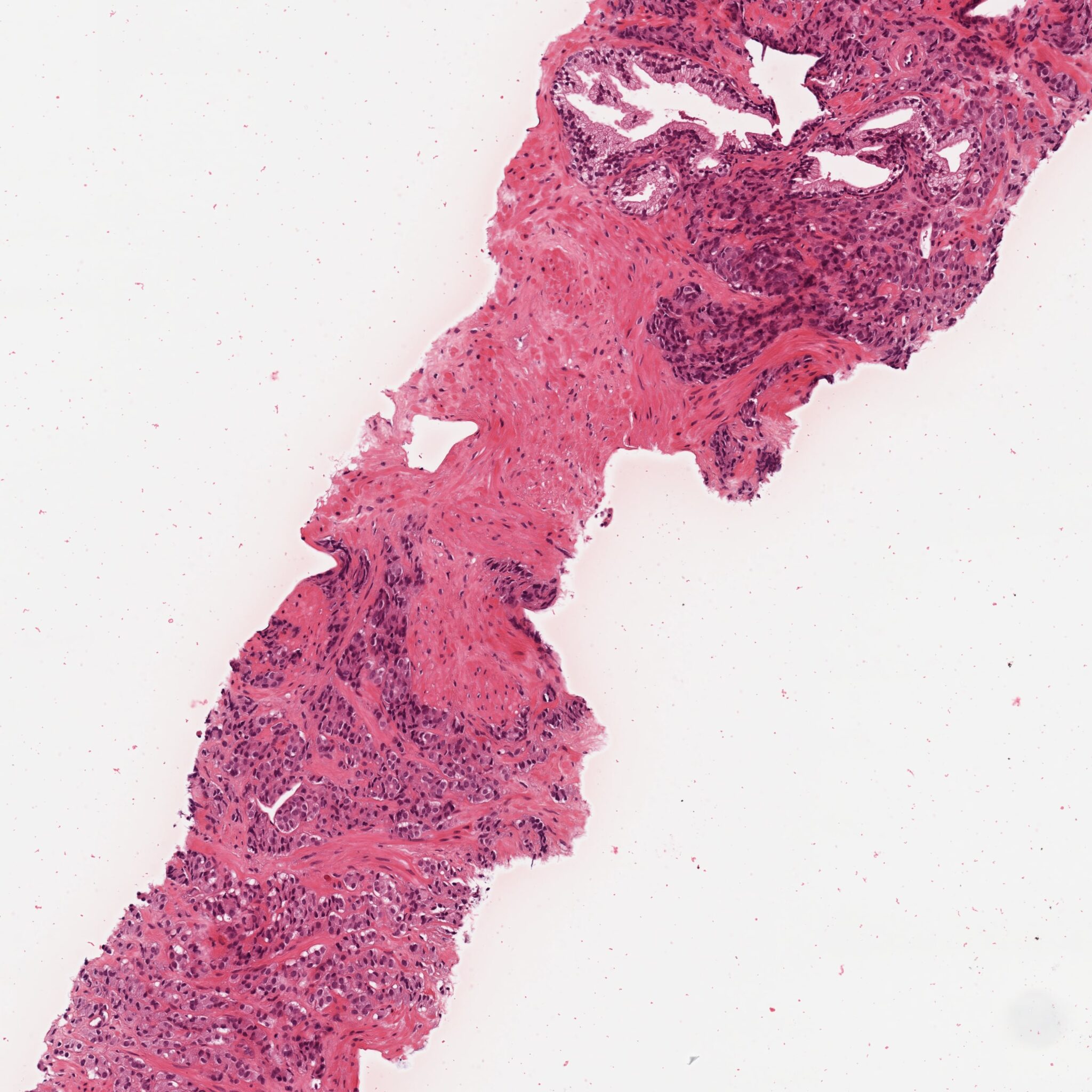
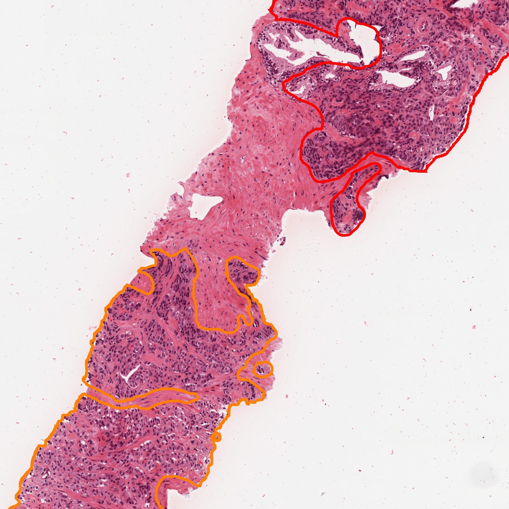
Image of prostate tissue stained with hematoxylin and eosin, original (left) and processed with ASSIST (right)
ASSIST Breast
ASSIST* Breast uses AI models to locate and characterize cell nuclei in digital images of breast tissue with immunohistochemical staining for the following biomarkers: ER, Ki67, PgR.
- It automatically segments cell nuclei, classifying them into marker-positive and marker-negative cell nuclei. Positive nuclei are classified as weak-, moderate-, and strong-positive.
- It provides segmentations, excluding non-representative areas or areas containing artefacts.
- It extracts quantitative parameters relevant to the diagnostic process, such as cell nuclei count and Positivity Ratio.
- It automatically generates standardised reports, providing pathologist with objective and reproducible support for diagnostic evaluation and characterization of breast tissue.
*Device undergoing certification according to Regulation (EU) 2017/746 (IVDR).
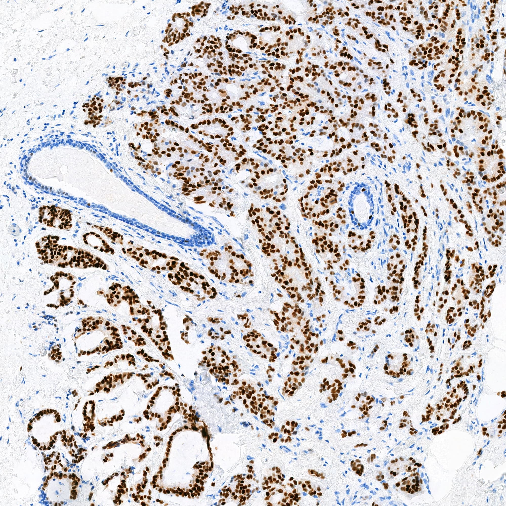

Image of breast tissue with immunohistochemical staining (ER), original (left) and processed with ASSIST (right)
Don’t have your credentials yet?
Mail us and you will discover all the potential of ASSIST