The challenge of Anatomic Pathology
According to current projections, new cancer cases are expected to increase by more than 60% by 2040, from 18.1 million new cases in 2018 to 29.4 million cases in 2040.

The medical field that studies cancer is Anatomic Pathology, which provides diagnoses from the analysis of organic tissues. Biological tissue can be examined in the form of histological slides, which is a complex and methodological activity that requires extreme precision.
Recently, histological slides have become digital thanks to technological developments. With a digital slide, it is possible to observe the biological tissue using a standard computer monitor.
Clinical pathologists daily analyze dozens of physical and/or digital slides to provide an accurate diagnosis.


Visual feedback of the human eye and brain has, by its very nature, limits.
Tissue analysis currently relies on visual feedback of the human eye and brain which, by their nature, have limitations. Ancient sceptics (Pyrrhon, Timon, Arcesilaus, Carneas… yeah Carneas, who was that?) had already observed that, since our senses can sometimes deceive us, it could be possible that they always deceive us and we can never be absolutely sure that we are not mistaken. Our sense of sight is particularly unreliable, as can be clearly seen from the ingenious optical experiments developed by Gestalt psychologists. According to this school of thought, our visual perceptions tend to organise data from the nervous system according to certain organisational principles and pre-established patterns (proximity, similarity, figure-background, etc.). These ‘interpretations’ of perceptual data give rise to a series of optical illusions which we must be aware of.
SIMILARITY AND PROXIMITY
Within an image, elements that are similar to each other are grouped together by our brain and perceived as one element. This “similarity” may be by either shape, color, size, or position.When the balls are all at equal distances, you see them as forming the shape of a square, but if you create a space between them, then you see three columns of balls.
COLOUR PERCEPTION
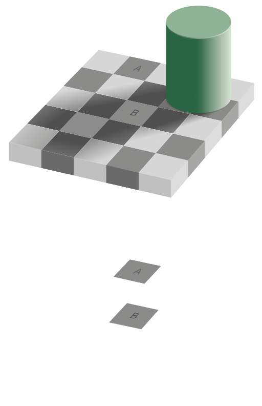
Colour is not solely a property of the observed object, but the combination of light, visual perception and the object properties. The three aspects that affect the perception of colour are hue, clarity and saturation, and how they are combined can give rise to surprising illusions.Squares A and B are the exact same colour. It seems incredible, and yet… our brain is tricked by lights and shadows.
DIMENSIONS PERCEPTION
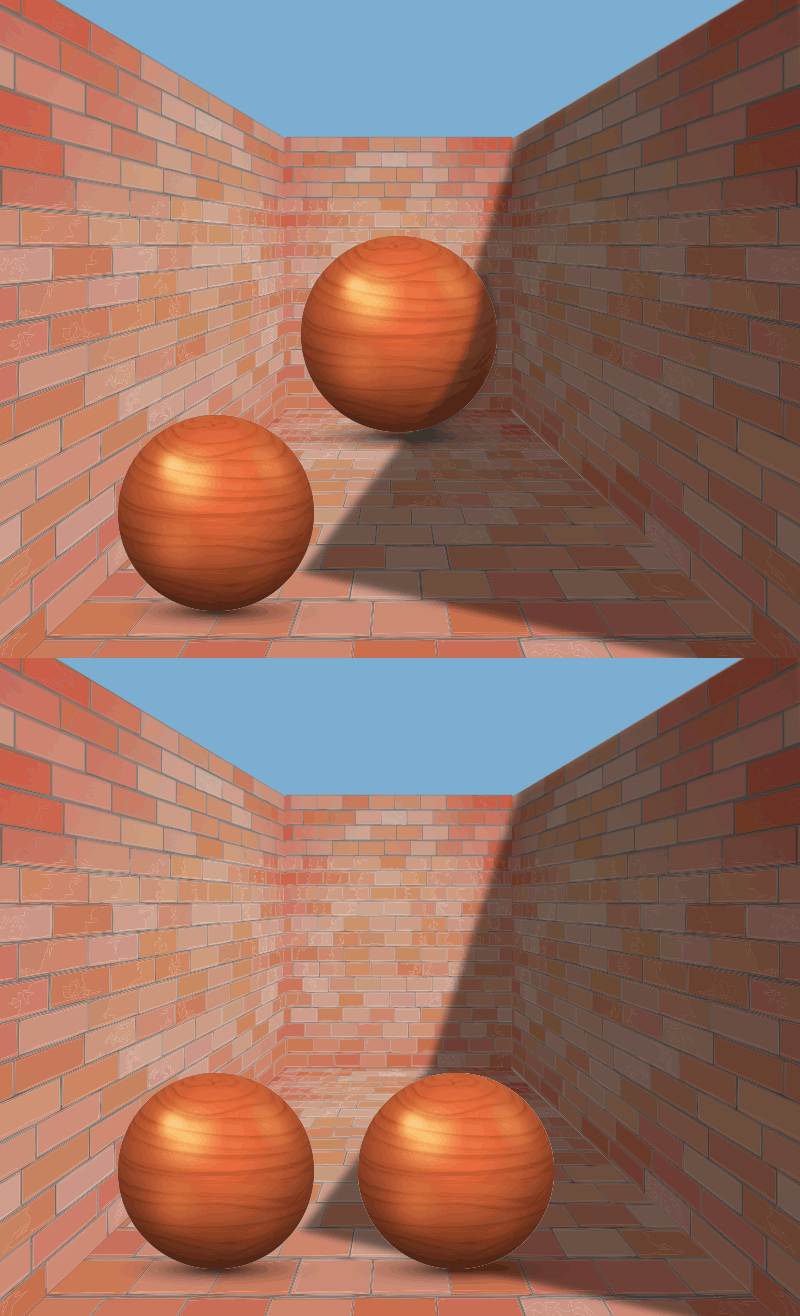
The perceived size of two or more objects depends on their location in space and on colors, patterns, etc.The way perspective is drawn in the picture makes the sphere on the right appear bigger, but both spheres are exactly the same size.
The Pathologist-Machine duo for a faster and more precise diagnosis.
This is all the more true in medical fields and, therefore, also in Anatomic Pathology. Due to the nature of their work, pathologists cannot afford to be hindered by such dynamic illusions…
AEQUIP is a possible solution to these problems: a technological support to the pathologist in all diagnostic phases, to help him/her provide increasingly precise and accurate diagnoses and minimizing the possibility of errors, gestalt or not.
AEQUIP is based on the most innovative technologies available today: Artificial Intelligence, Machine Learning and Deep Learning. Since these algorithms are highly sensitive to the quality of the input data, we combine these technologies with mathematical algorithms to ensure greater control over their performance and refine the output results.
A complex system that we define “M.D.I.”. Mathematical-Driven Intelligence.
Workflow


The process is not automatic, so it is subject to:
• high stain variability
• suboptimal staining

The presence of different scanner models results in increased staining variability of the scanned slides in different centres.


The diagnostic process is hampered by several factors:
•Increase in cancer cases
• Increase in details required for diagnosis
• Qualitative rather than quantitative analysis


