STAINS
STAndardIzation & Normalization of histological Slides
The quality of histology slides is often suboptimal, which can result in delayed diagnoses and lower diagnostic accuracy. STAINS (STAndardIzation & Normalization of histological Slides) can improve the quality of the digital images available to the pathologist.
Typical critical issues in histopathological diagnosis
Histopathological diagnosis plays a crucial role in medical practice, especially in the analysis of tumors. Often, visual inspection of samples can be hampered by suboptimal slide quality and variability in staining, which can negatively impact the process in terms of both accuracy and time. These problems are exacerbated when placed in the context of increasing pressure on laboratory operators and pathologists due to the rise in cancer cases and the increasing detail required in reports. Similarly, digital images with high staining variability can decrease the performance of the entire Pathology department workflow.
STAINS is a solution
An automatic algorithm designed specifically to address these issues, STAINS is able to digitally recalibrate the color profile of a poor-quality histological image to make it similar to the optimal one chosen by the pathologist, reducing the variability introduced during the staining and scanning process of the samples.
During the reporting process, STAINS allows the pathologist to observe the sample with optimal staining, aiding in the diagnosis process.
STAINS is available for the following stains and tissues:
- Hematoxylin and Eosin (H&E) for prostate, lung, liver, breast, colon, stomach, and uterus tissues – In vitro diagnostic medical device – IVD, according to Regulation (EU) 2017/746 (IVDR);
- IHC staining with the following markers Ki67, PGR, HER2, and ER for breast tissue – In vitro diagnostic medical device – IVD, according to Regulation (EU) 2017/746 (IVDR);
- Special PAS staining for kidney, lung, and liver tissues*;
- Special trichrome staining (Masson-green) for kidney, myocardium, and liver tissues*.
STAINS product generates a new normalized digital slide without directly modifying the original slide.
* Device for Research Use Only (RUO)
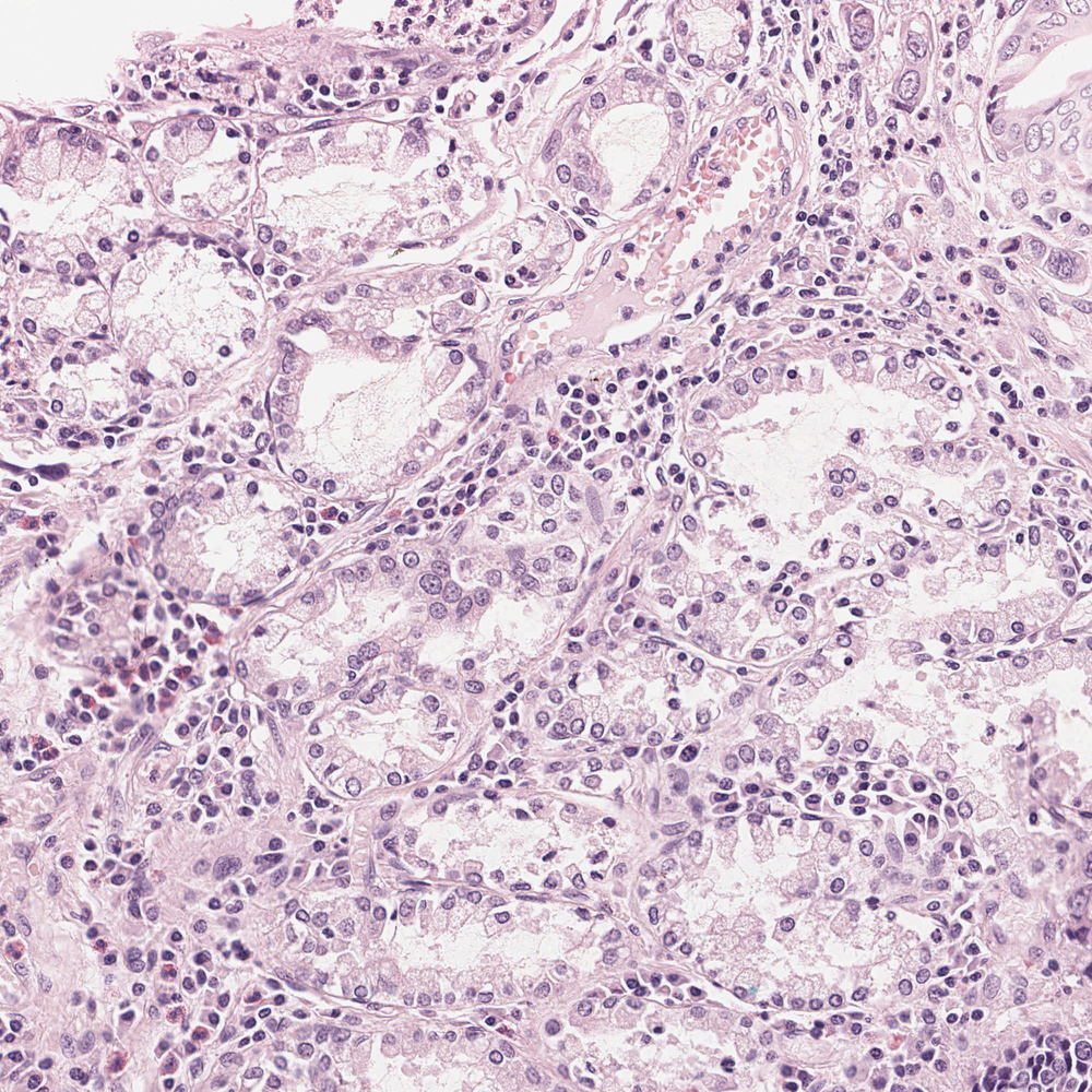
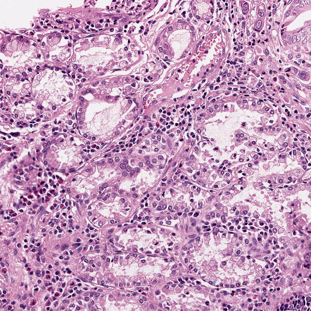
Image of stomach tissue stained with hematoxylin and eosin, original (left) and normalised with STAINS (right).
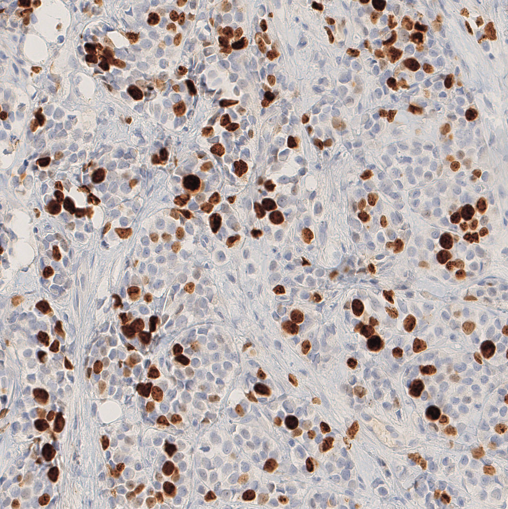
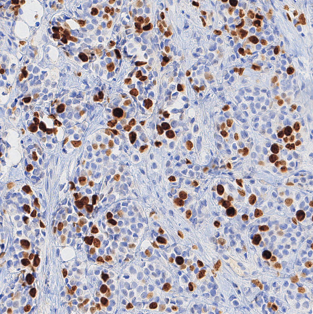
Image of breast tissue with immunohistochemical staining (Ki67), original (left) and normalised with STAINS (right).
Don’t have your credentials yet?
Mail us and you will discover all the potential of STAINS